Figure 3.

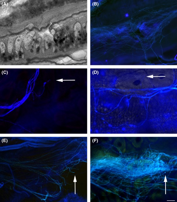
Pollen tube growth. (A and B) Pollen tube growth from crossings between Epidendrum fulgens × E. fulgens as a positive control – (A) light microscopy showing ovules and (B) fluorescent microscopy showing the pollen tube reaching the ovule level. (C and D) Pollen tube growth from crossings between E. puniceoluteum × hybrid (pollen receptor × donor) showing timid pollen tube germination reaching an ovule level. (E and F) Hybrid × E. puniceoluteum (pollen receptor × donor) showing pollen tubes reaching hybrid ovule level (arrows). Pollen tubes are in blue, acquired by fluorescence microscopy, and ovule photos were acquired by light contrast microscopy. (D–F) Merged images from fluorescence (pollen tube) and light microscopy (ovule). (A and C) Pollen tubes observed by fluorescence microscopy. Scale bar in (F) indicates 10 μm.
