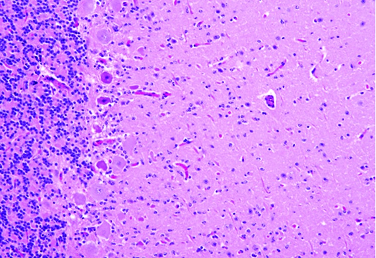
Figure 4.
Cerebellum of a yearling steer with encephalomyelitis (animal 1). Note the selective extensive acute necrosis and degeneration of Purkinje cells. Numerous necrotic dendritic spheroids in the molecular layer with a cellular proliferation of Bergmann glia and of microgliosis. Hematoxylin and eosin stain. Original magnification ×400.

