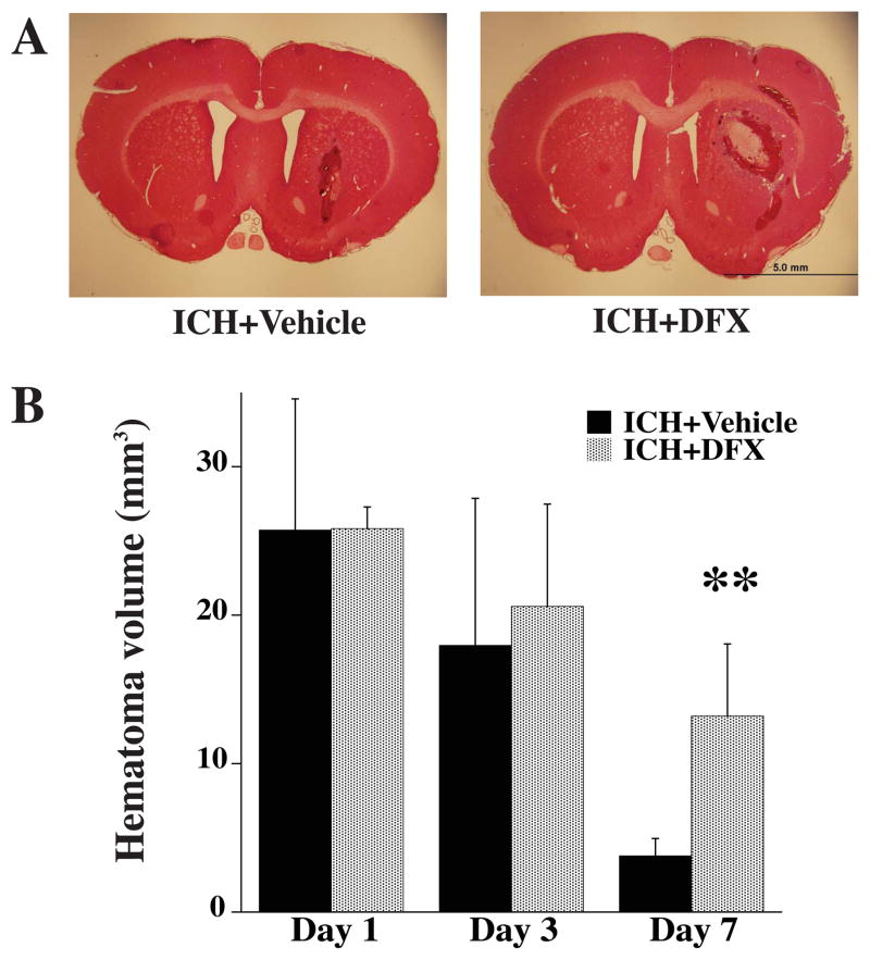
Figure 4.
A) Coronal sections (hematoxylin and eosin staining) from brains 7 days after ICH in vehicle and DFX-treated groups. B) Bar graph showing hematoma volume 1, 3 and 7 days after ICH in vehicle- and DFX-treated groups. Values are means±SD. n=4 rats per group, **P<0.01 vs. ICH+vehicle group. Scale bar = 5.0 mm.

