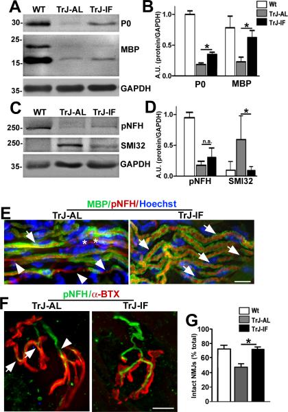
Figure 6.
Intermittent fasting (IF) improves distal nerve myelination and neuromuscular junction (NMJ) innervation. (A, C) Whole soleus muscle lysates (40 μg/lane) were analyzed with anti-myelin (myelin basic protein (MBP] and protein zero (P0]), and anti-neurofilament (pNFH and hypo-phosphorylated NFH (SMI32]) antibodies. Glyceraldehyde 3-phosphate dehydrogenase (GAPDH) is shown as a loading control. (B, D) Semiquantitative analyses of 4 independent blots. There is a significant increase in the levels of MBP and P0 in samples from intermittent fasted Trembler J (TrJ-IF), vs. ad libitum (AL)-fed littermates (TrJ-AL) (Student t-test, *p < 0.05) (B). The increased expression of pNFH in TrJ mice on the IF regimen (TrJ-IF) vs. TrJ-AL is not significant but the levels of SMI32-reactive proteins is significantly reduced in IF-fed mice (D). (E) Sections of soleus muscles from TrJ-AL and TrJ-IF mice were co-immunolabeled with anti-MBP (green) and anti-pNFH (red) antibodies. MBP-positive myelin sheaths (arrows) and demyelinated axon segments (arrowheads) are denoted. Unmyelinated fibers are marked with asterisks. Nuclei are stained with Hoechst dye (blue). (F) Sections of soleus muscles from TrJ-AL and TrJ-IF mice were co-labeled with an anti-NFH antibody (green) and α-BTX (red). Thinning (arrows) and axonal retraction (arrowheads) at the NMJs are marked in the NMJ from the TrJ-IF soleus. Scale bars: 10 μm (E, F). (G) Quantification of intact NMJs in soleus samples from the indicated groups. (Student t-test, *p < 0.05). n = 3 to 4 mice per genotype and diet regimen.

