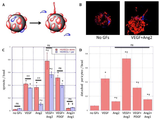
Figure 2.
Schematic (A) and microscopy (B) images of R18-labeled endothelial cell (red) sprouting and CMFDA-labeled pericyte (blue) detachment from microcarriers in response to growth factor signals. (C) Endothelial cell sprouting from microcarriers over three days with EC (red bars) or EC/pericytes (blue bars) cultures in fibrin in response to VEGF (50ng/mL), Ang2 (250ng/mL), combined VEGF(50ng/mL) and Ang2(250ng/mL, VA2), combined VEGF and PDGF(both 50ng/mL, VP) or combined Ang2 and Ang1 (both 250ng/mL, Ang1). (D) Pericyte migration away from endothelium on microcarriers with EC/pericyte co-cultures in response to conditions specified in C. *: statistically significant relative to no GF control. †: statistically significant relative to VEGF; ‡: statistically significant relative to Ang2; **: statistically significant between presence and absence of pericytes; ns: not significant by Student’s T-test. Data represent mean and S.E.M.

