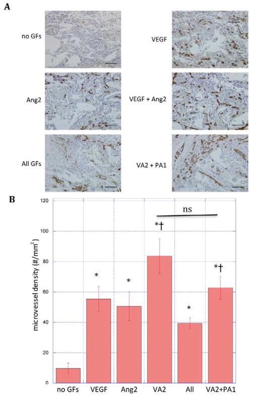
Figure 4.
(A) Photomicrographs of hematoxylin and CD31 stained section of scaffolds and (B) quantification of microvessel density in PLG scaffolds subcutaneously implanted in mice for two weeks. Scaffolds (n = 3) containing no growth factors (no GFs), rapidly releasing VEGF, Ang2, VEGF and Ang2 simultaneously (VA2), VEGF, Ang2, PDGF and Ang1 all simultaneously (all GF), or rapidly releasing VEGF and Ang2 followed by delayed release of PDGF and Ang1 (VA2+PA1). Data represent mean and S.E.M.
*: statistically significant relative to no GFs; † statistically significant relative to All GFs; ns: not significant by Student’s T-test. scale bar: 100μm

