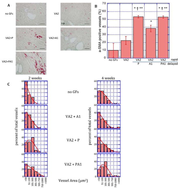
Figure 5.
Delayed release of pro-maturation factors increases mural cell association and vascular remodeling. A: Photomicrographs of α-SMA-stained sections of scaffolds implanted subcutaneously releasing no growth factors (no GFs), VEGF/Ang2 (VA2) alone or followed by a delayed release of PDGF (P), Ang1 (A1), or both (PA1) at two weeks. B: Quantification of the percentage of blood vessels associating with smooth muscle cells determined by α-smooth muscle actin staining of tissue sections from subcutaneously-implanted scaffolds C: Cross-sectional area of vessels formed by rapid delivery of VEGF/Ang2 (VA2) followed by delayed release of PDGF (P), Ang1 (A1), or both (PA1) at two and four weeks. Values represent mean and S.E.M.
*, †, ** Denote p<0.05 relative to blank, VA2, and VA2+A1, respectively by Student’s T-test. Scale bar = 100μm

