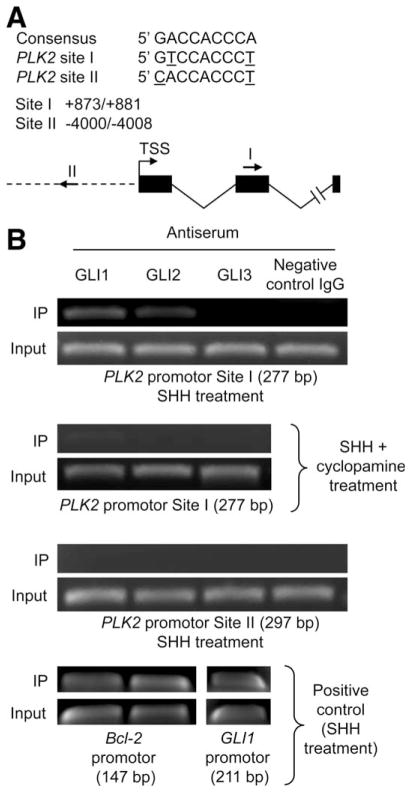
Fig. 3.
GLI1 and GLI2 bind to a predicted GLI-binding site in the PLK2 promoter region. (A) Two putative GLI-binding sites that contain two mismatches, compared to the consensus sequence, were identified in the PLK2 promoter region. Nucleotide positions were counted from the transcription start site (TSS). Positions and directions of both potential binding sites are illustrated by arrows (I and II). (B) KMCH-1 cells treated with rhSHH (500 ng/mL, 5 hours)±cyclopamine (10μM, 5 hours) were employed for this study. ChIP using antiserum to GLI1, GLI2, and GLI3 or a sheep negative control IgG was performed, followed by PCR using primer flanking site I (277 bp) or II (297 bp) within the PLK2 promoter region. As positive controls, ChIP was performed using primers flanking the Bcl-2 promoter GLI-binding sites (GLI1 and GLI2; 147 bp) or primers flanking the GLI3-binding site within the GLI1 promoter (211 bp).

