. 2013 Sep;15(3):263–278. doi: 10.31887/DCNS.2013.15.3/goz

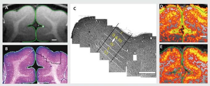
Figure 1. Coregistration of magnetic resonance (MR) image with the corresponding cortical tissue: Following the MR imaging session, the animals were sacrificed and the cortical tissue was stained for cytoarchitectonic structures, (a) A high-resolution MRI. (b) Low-magnification photograph of the corresponding Nissl stain (histology) of the same region, (c) A mosaic reconstruction of high magnified Nissl-stained images (right hemisphere) of cortical tissue corresponding to the imaged plane (outlined in panel b). White lines: the anatomically defined borders between cortical layers, (d) and (e) High-resolution blood oxygen level-dependent and cerebral blood volume functional MRI (0.15x0.15 mm2) maps for a 40-s drifting bars stimuli. Signal changes were confined to the primary visual areas and closely followed the gray matter contour. In both maps, a spatially defined band of elevated signal changes was centered over layer IV as defined anatomically in panel c. Adapted from ref 44: Harel N, Lin J, Moeller S, Ugurbil K, Yacoub E. Combined imaging-histological study of cortical laminar specificity of fMRI signals. Neuroimage. 2006;29:879-887. Copyright © Elsevier 2006.

Figure 1.