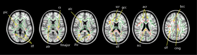
Figure 5. Differences in white matter integrity in autism. Tract-based spatial statistics revealed regions of reduced fractional anisotropy in children with autism spectrum disorder compared with the typically developing group. Red color symbolizes significant voxels at P<.05 (corrected for multiple comparisons at cluster level). Mean skeleton of detected fiber tracts is overlaid in green on standard T1-weighted anatomical image), ilf, inferior longitudinal fasciculus; ifo, inferior fronto-occipital fasciculus; slf, superior longitudinal fasciculus; cs, corticospinal tract; cing, cingulum; bcc, body of corpus callosum; gcc, genu of corpus callosum; sec, splenium of corpus callosum; aic, anterior internal capsule; pic, posterior internal capsule; fmajor, forceps major; acr, anterior corona radiata; scr, superior corona radiata; atr, anterior thalamic radiation Reproduced from ref 79: Shukla DK, Keehn B, Lincoln AJ, Muller RA. White matter compromise of callosal and subcortical fiber tracts in children with autism spectrum disorder: a diffusion tensor imaging study. J Am Acad Child Adolesc Psychiatry. 2010;49:1269-1278.e2. Copyright © Elsevier 2010.