. 2013 Sep;15(3):359–384. doi: 10.31887/DCNS.2013.15.3/edennis

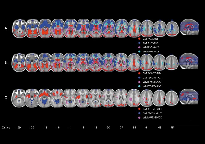
Figure 7. Differences in regional brain volume in fragile X. A: Regions showing significant differences in regional gray matter (GM) volume and white matter (WM) volume between fragile X syndrome (FXS) and idiopathic autism (iAUT) (panel A), FXS and typically developing (TD) and idiopathic developmentally delayed (DD) controls (panel B), and iAUT and TD/DD controls (panel C). The left side shows the right hemisphere. The statistical threshold is set at P=0.01 , familywise error cluster-level corrected. Montreal Neurological Institute coordinates. Adapted from ref 118: Hoeft F, Walter E, Lightbody A, et al. Neuroanatomies! differences in toddler boys with fragile X syndrome and idiopathic autism. Arch Gen Psychiatry. 2011;68:295-305. Copyright © American Medical Association 2011.

Figure 7.