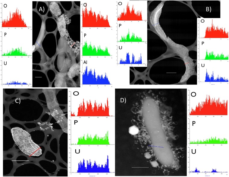
Fig 5.
XEDS of the wild type (A and B), the ΔpilA mutant (B), or the ΔBESTZ mutant (C) respiring U(VI). High-angle annular dark-field STEM images of areas of freeze-dried cryo-TEM grids are shown. The “spider web-like” pattern supporting the cells is the lacey carbon support. The scattering from metal aggregates and gold beads appears intensely bright. The red and blue lines indicate the line scanned by the probe. Scale bar, 500 nm. Side panels show X-ray counts of the main elements along the scanned line. The units in the line scans are nm on the x axis and X-ray counts on the y axis. For O (oxygen), P (phosphorus), and Al (aluminum), it is the number of X-ray counts in their K alpha peaks, and for U, it is the number of counts in the L alpha peak. Uranium counts are significantly above the background level.

