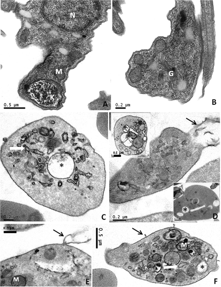
Fig 2.
Transmission electron microscopy analysis of cynaropicrin's effects on bloodstream trypomastigotes. BT were left untreated (A and B) or exposed to this STL (EC50/24 h) for 2 h (C to F). Untreated parasites displayed typical morphology, while cynaropicrin-treated parasites showed vacuolization (*), swelling of the mitochondrion and endoplasmic reticulum (white arrows), and plasma membrane shedding (black arrows). M, mitochondrion; G, Golgi complex; N, nucleus.

