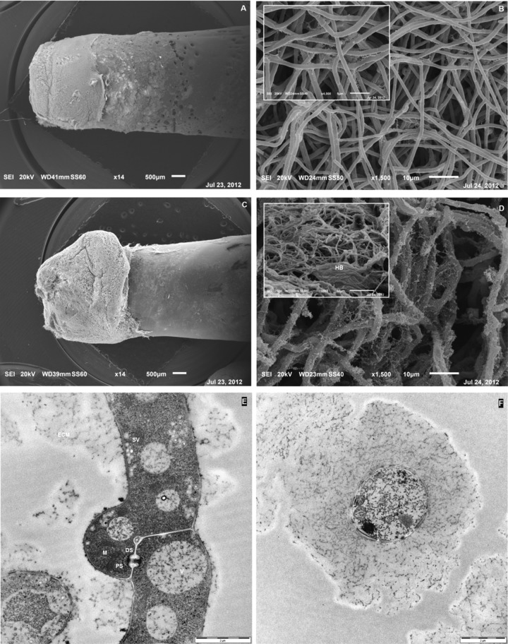
Fig 3.
(A to D) SEM micrographs of P. ostreatus biofilms grown on the HA-P&G system and LB medium for 4 days (A and B) and 7 days (C and D) at 30°C (150 rpm). Insets show details at higher magnifications. (E and F) TEM micrographs of longitudinal and cross-view sections of a biofilm's constituent hypha, respectively. Abbreviations: DS, dolipore septum; ECM, extracellular matrix; HB, hyphal bundle; PS, parenthosome; SV, secretory vesicles; V, vacuoles.

