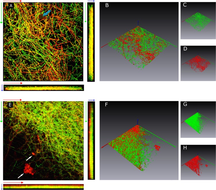
Fig 4.
CLSM images of 4-day-old (A to D) and 7-day-old (E to H) P. ostreatus biofilms grown on the HA-P&G device under orbital shaking (150 rpm) at 30°C and sequentially stained with concanavalin A conjugated with Texas Red (red emission) and Calcofluor White Stain M2R (green emission). Images A and E are horizontal (xy) and vertical side (xz and yz) views of three-dimensional reconstructed images of 4- and 7-day-old biofilms obtained by volume rendering, respectively, as described by Harrison et al. (32). Isosurfaces of 4- and 7-day-old biofilms are shown in panels B to D and F to H, respectively. Each image represents an area of 375 by 375 μm. The light blue and white arrows in panels A and E indicate a hydroxyapatite granule and ECM aggregates, respectively. The x, y, and z axes are coded in red, green, and blue, respectively. The direction of the blue arrow (z axis) is oriented toward the outermost biofilm layers.

