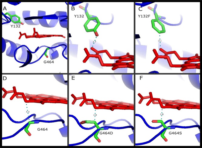
Fig 1.
Positions of the point mutations of Candida tropicalis Erg11p analyzed according to the homology models generated in this study. (A, B, and D) Wild-type Candida tropicalis Erg11p (Y132 and G464). (C, E and F) Positions of the mutations analyzed in the protein: Y132F, G464D, and G464S, respectively. The residues studied are shown in stick representation (green, carbon atoms; red, oxygen atoms; blue, nitrogen atoms). Heme is represented in red. The distance between the heme and the closest atom of the amino acid studied is indicated by a dashed green line and a number giving the distance in daltons. The crystal structure of human lanosterol 14α-demethylase (HS-CYP51) in complex with ketoconazole (Protein Data Bank code 3LD6) was used as a template. Y, tyrosine; F, phenylalanine; G, glycine; D, aspartic acid; S, serine.

