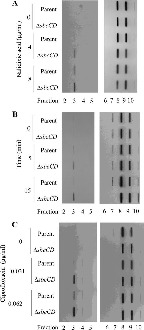
Fig 3.

Increased levels of covalently bound gyrase in the ΔsbcCD mutant compared to parental strain. (A) Exponentially growing cultures of E. coli parental strain BW27784 and its ΔsbcCD mutant derivative were incubated for 30 min in the absence or presence of 4 μg/ml (1× MIC) or 8 μg/ml (2× MIC) of nalidixic acid. Cells were then lysed in the presence of a detergent, and gyrase-DNA complexes were separated from free gyrase by sedimentation in a CsCl density gradient. Gradients were fractionated from bottom to top and loaded on a slot blot. Slot blots were probed with an anti-GyrA antibody. (Left) CsCl density gradient fractions 2 to 5, collected from the bottom portion of the gradient (higher density). (Right) CsCl density gradient fractions 6 to 10, collected from the top portion of the gradient (lower density). (B) The parent and the ΔsbcCD mutant were grown to exponential phase, and an aliquot was collected immediately before the addition of nalidixic acid to a final concentration of 8 μg/ml (2× MIC) (0 min). After 5 and 15 min of incubation with nalidixic acid, more aliquots were taken. Gyrase-DNA complexes were isolated as described for panel A. (C) The parent and the ΔsbcCD mutant were incubated for 5 min in the absence or presence of 0.031 μg/ml (1× MIC) or 0.062 μg/ml (2× MIC) of ciprofloxacin. Gyrase-DNA complexes were isolated as described for panel A.
