
Fig 2.
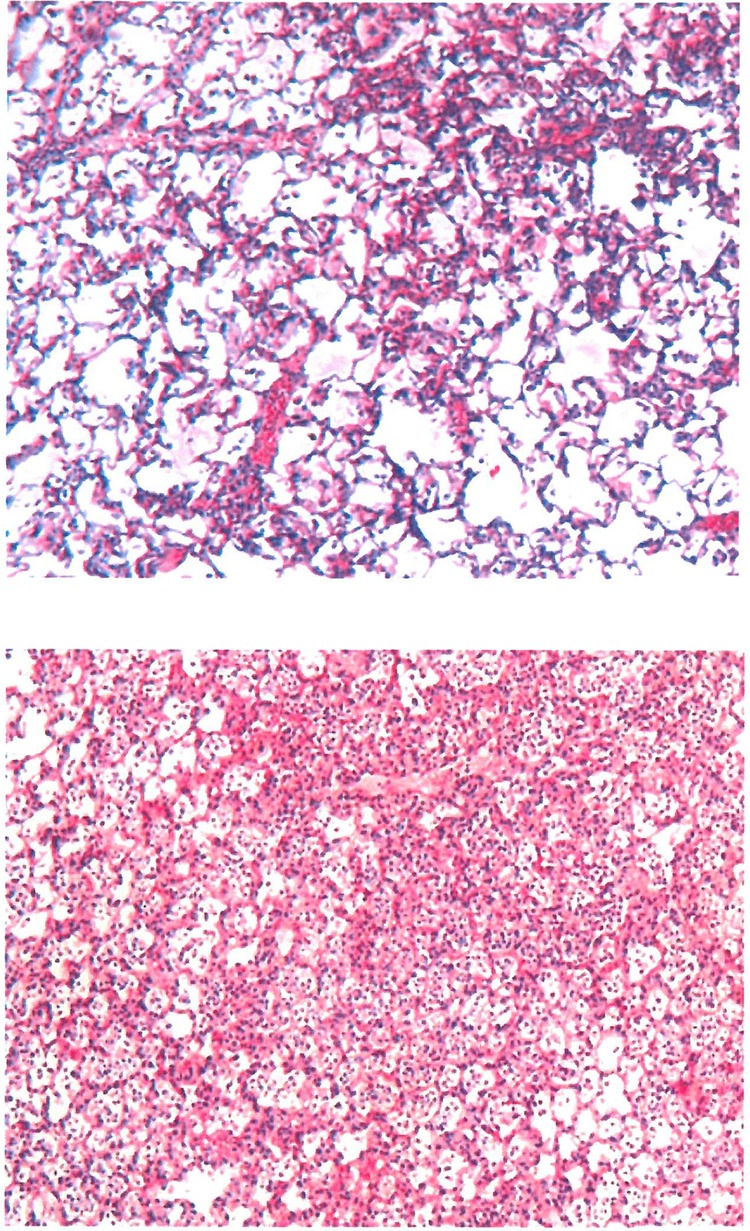
Pathological mapping of lung tissue samples representative of the ABCR and ABCS groups. Giemsa staining and a magnification of ×50 were used. ABCR lung tissue showed fewer confluent lesions of bronchopneumonia with areas of preserved lung architecture, in contrast to ABCS lung tissue.