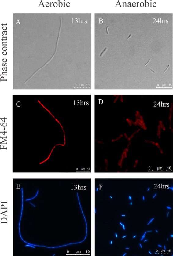
Fig 2.

Cell morphology of E. coli strain TOP10 after induction of LL-37 expression under aerobic and anaerobic growth conditions. The cell morphologies of E. coli strains harboring pHERD30T-LL37 were detected by phase-contrast microscopy after induction with 0.2% arabinose under aerobic growth for 13 h (A) and anaerobic growth for 24 h (B). Cell membranes (C and D) were stained with FM 4-64 and DNA (E and F) with DAPI and visualized by epifluorescence microscopy.
