Fig 3.
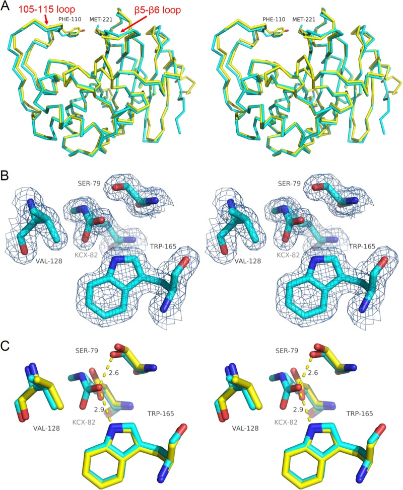
Comparison of OXA-23 and OXA-24 structures. (A) Structural alignment of the α-carbon traces of OXA-23 (cyan) and OXA-24 (yellow; PDB accession no. 3PAE). Also shown are residues that form the bridge over the top of the active site (F110/M221 in OXA-23 and Y112/M223 in OXA-24). (B) 2Fo-Fc electron density maps of key active-site residues of OXA-23 contoured to 1.0 σ showing full carboxylation of K82. (C) Structural alignment of OXA-23 (cyan) and OXA-24 (yellow) active-site residues showing the high degree of overlap (OXA-24 structure 3PAE contains a K84D substitution).

