
Fig 2.
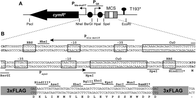
(A) Organization of cymR*, PQ5, and the multiple-cloning site (MCS) on plasmid pQF (not to scale). Restriction sites flanking important features are indicated. T193* denotes a putative transcriptional terminator derived from Sphingomonas Fr1 (see the supplemental material). Gray boxes in the MCS indicate 3×FLAG tags and black circles CuO sites. (B) Nucleotide sequence of the PQ5 promoter region and the MCS. cymR* and MCS start codons are in bold. Ribosome binding sites (RBS), operator sequences (CuO), and −35 and −10 promoter elements are boxed. Core promoter sequences of PQ5 (Psyn2) and the promoter driving cymR* expression (Pbla-mut1T) are indicated by arrows. Translation of the MCS is indicated below the nucleotide sequence. All restriction sites shown are unique in pQF, except those marked with an asterisk. The 3×FLAG tag (peptide sequence DYKDHDGDYKDHDIDYKDDDDK) is shown schematically.