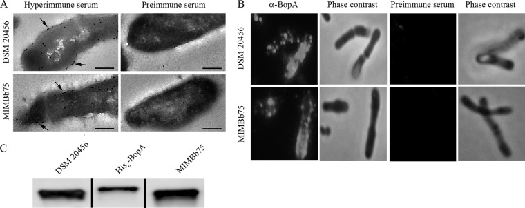
Fig 2.

Subcellular localization of BopA in B. bifidum DSM20456 and MIMBb75. (A) Immunoelectron microscopy images of the localization of BopA in B. bifidum cells. Detection was done on thin sections by using anti-BopA (hyperimmune serum) and protein A-gold particles (pAp). The arrows indicate the pAp binding to the cell surface. Preimmune serum labeling of cells was used as a negative control. Bar, 200 nm. (B) Immunofluorescence staining of B. bifidum DSM20456 and MIMBb75 cells with anti-BopA and Alexa-488-conjugated secondary IgG. Preimmune serum was used as a negative control. Phase-contrast images of the same microscopic fields are shown on the right. (C) Western blotting of BopA in the cell wall extracts of B. bifidum DSM20456 and MIMBb75. His6-BopA (5 ng) is shown for comparison.
