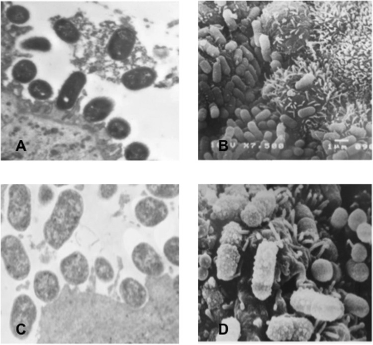
Fig 1.

(A) Transmission electron microscopy (TEM) of human duodenal mucosa infected with an O26:H11 strain (1551-3/85), showing attachment-effacement lesions comprised of closely attached bacteria accompanied by alterations to the enterocyte cytoskeleton, evidenced by cupping, pedestal formation, accumulation of electron-dense material, and an absence of microvilli at sites of bacterial attachment. Magnification, ×12,500. (B) Scanning electron microscopy (SEM) of human duodenal mucosa infected with an O26:H11 strain (1551-3/85) confirms the occurrence of bacterial colonization of the mucosal surface. Magnification, ×9,940. (C and D) TEM and SEM views of human duodenal mucosa ex vivo infected with an O26:H11 strain (0791-1/85), showing bacterial adhesion but without true pedestal formation. Magnification, ×12,120 (C) and ×11,310 (D).
