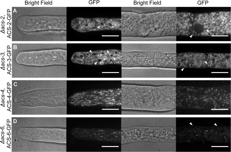
Fig 4.
Localization of ACS-GFP fusion proteins. ACS-GFP fusions were expressed in the background strain of the respective Δacs strain. Bright-field and GPF fluorescence images were obtaining by spinning disc confocal microscopy (see Materials and Methods). The two columns of panels on the left show localization in the tip region of a hypha, while the two right columns show localization in subapical regions. Arrows indicate putative endoplasmic reticulum (ER) localization for ACS-2–GFP and ER, plasma membrane, and septal localization for ACS-3–GFP. For ACS-6–GFP, arrows show plasma membrane localization. Bars = 10 μm.

