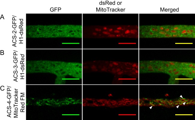
Fig 5.

Colocalization of ACS proteins with fluorescent markers for different cellular structures. (A) Forced heterokaryon of Δacs-2 ACS-2–sGFP plus Δrid H1-dsRed strains. (B) Forced heterokaryon of Δacs-3 ACS-3–sGFP plus Δrid H1-dsRed strains. ACS-2–GFP and ACS-3–GFP localizes to the membrane surrounding the nuclei, as indicated by the H1-dsRed fluorescence, indicating endoplasmic reticulum localization. (C) Δacs-4 strain ACS-4–GFP colocalizes with the mitochondria, as indicated by MitoTracker Red FM dye. Fluorescence images were obtaining by spinning disc confocal microscopy (see Materials and Methods). Bars = 10 μm.
