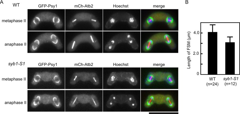
Fig 6.

(A) Aberrant assembly of the FSM in syb1-S1 cells. Wild-type (TY235) and syb1-S1 (TY234) strains expressing GFP-Psy1 and mCherry-tagged Atb2 (α-tubulin) were sporulated on MEA medium at 28°C for 1 day. Chromosomal DNA was stained with Hoechst 33342 and analyzed by fluorescence microscopy. Bar, 10 μm. (B) Development of the FSM in syb1-S1 cells. The contour length of FSMs in wild-type (TY235) and syb1-S1 (TY234) cells at late meiosis II was measured by Image J software.
