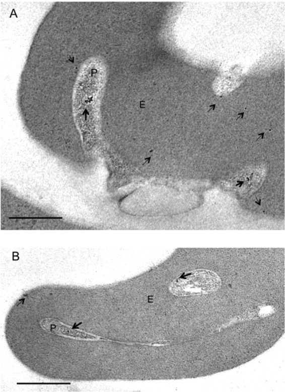
Fig 6.

Immunoelectron microscopy indicates the location of PfShelph2 in early-ring-infected erythrocytes. Invasion events and early rings were captured, and thin sections were probed for the PfShelph2 location as described in Materials and Methods. Gold particles (10 nm) indicate the location of PfShelph2. Dark and light arrows, respectively, mark PfShelph in the parasite and erythrocyte. (A) Parasite (P) during entry into the erythrocyte (E) and in the process of ring formation. (B) Newly formed intracellular ring stage parasite (P) in erythrocyte (E). Scale bars, 0.5 um. Control sections probed with secondary antibodies alone showed no gold particles (see Fig. S6 in the supplemental material).
