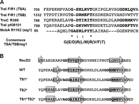
Fig 2.

(A) ClustalO alignment of TSs determined for R1/F-TraI and R1162-MobA against the whole R388-TrwC and pKM101-TraI sequences. The asterisks indicate positions that have a single, fully conserved residue; the colons indicate conservation between groups of strongly similar properties. (B) The protein secondary-structure prediction was obtained using Phyre2 for RecD2; wild-type TrwC; and TrwC variants TS1*, TS2*, and TS1* plus TS2*. To simplify the results, the consensus secondary structures are represented by showing the amino acid sequences and the β-strands (arrows) or α-helices (cylinders).
