
Fig 7.
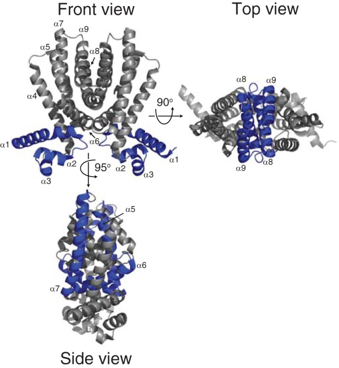
TFRs share nine conserved α helices. In the front view, the DNA-binding domain is made up of helices 1 to 3. In the side view, helices 5 to 7 in the ligand-binding domain form a central triangle. In the top view, helices 8 and 9 from each monomer form a four-helical bundle that makes up the dimer interface. The structure of Rha06780 (PDB ID 2NX4) is shown, as it shows a structure typical of TFRs (24).