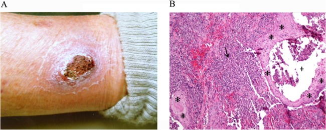
Fig 1.

(A) The cutaneous lesion from which Francisella sp. LA11-2445 was isolated. (B) Histopathology (100× magnification) of a fragmented punch biopsy specimen. The arrow marks a dense, mixed dermal infiltrate of neutrophils, histiocytes, and lymphocytes. An intraepidermal neutrophilic microabscess is shown (+). The asterisks (*) indicate pseudoepitheliomatous epidermal hyperplasia.
