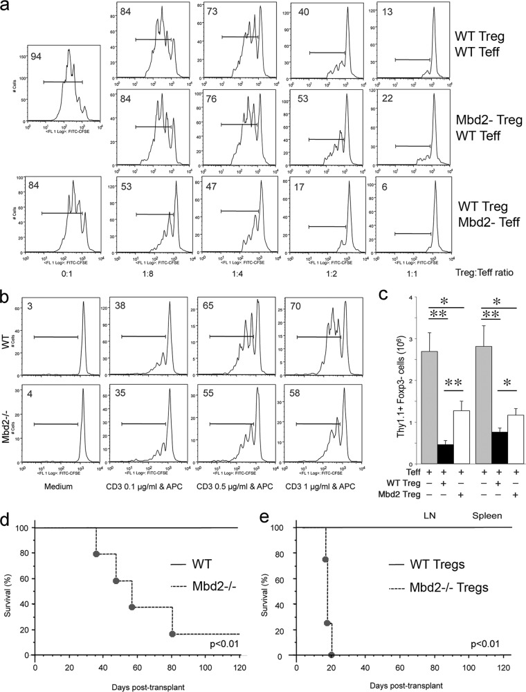
Fig 2.
Mbd2 deletion impairs Treg function in vitro and in vivo. (a) In vitro Treg suppression assays based on CFSE dilution by Teff cells proliferating in the presence of various ratios of WT or Mbd2−/− Treg cells; the proportion of proliferating cells is shown in each panel, and data are representative of 3 separate assays. (b) Mbd2−/− Teff cells display modestly impaired proliferative ability compared to WT Teff cells; the proportion of labeled cells is shown in each panel, and data are representative of 3 separate assays. (c) Mbd2−/− Treg cells displayed less ability than WT Treg cells to suppress the homeostatic expansion by 7 days of Thy1.1+ Foxp3− Teff cells that were adoptively transferred to Rag1−/− immunodeficient mice. Single-cell suspensions from LN or spleen samples were analyzed by flow cytometry for fluorescence-activated cell sorter (FACS) analyses (means ± SD, 4 mice/group). *, P < 0.05; **, P < 0.01 (for comparisons between groups, as shown). (d) Inability of a tolerance-inducing CD154 MAb/donor splenocyte transfusion protocol to prevent rejection in Mbd2−/− versus WT recipients of fully MHC-mismatched vascularized cardiac allografts (n = 5/group). (e) Differential survival of C57BL/6 cardiac allografts in BALB/c Rag1−/− immunodeficient hosts that were adoptively transferred with recipient Teff cells plus either WT or Mbd2−/− Treg cells (2:1 ratio of Teff to Treg cells, n = 4 mice/group).

