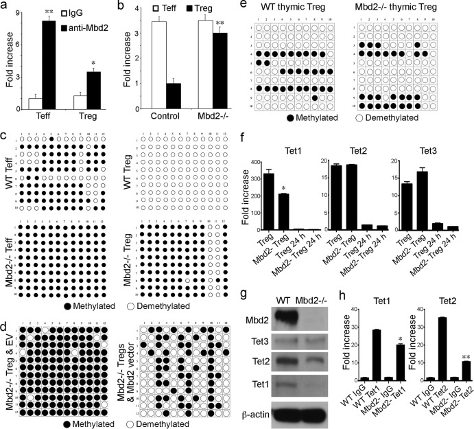
Fig 4.
Mbd2 associates with Foxp3 and affects Foxp3 methylation. (a) ChIP assay using anti-Mbd2 Ab to pull down Mbd2-associated chromatin, followed by Sybr real-time qPCR to detect the TSDR of Foxp3; data are shown as fold increase using purified Teff and Treg WT cells (*, P < 0.05, **, P < 0.01 [for anti-Mbd2 Ab versus control IgG]). (b) Methylation at the Foxp3 TSDR site was assessed using a methyl collector (Mbd2 protein) to pull down methylated DNA, followed by Sybr real-time qPCR; data are shown as fold increase using purified Teff and Treg cells (**, P < 0.01 [for Mbd2−/− versus WT Treg cells]). (c) Analysis of individual CpG sites within the Foxp3 TSDR by bisulfite conversion, cloning, and sequencing, using WT versus Mbd2−/− Teff and Treg cells. (d) Analysis of individual CpG sites within the Foxp3 TSDR by bisulfite conversion, cloning, and sequencing, using Mbd2−/− Treg cells retrovirally transduced with either empty vector (EV) control or vector containing Mbd2. (e) Bisulfite conversion, cloning, and sequencing showed that Mbd2−/− and WT thymic Treg cells had comparable levels of TSDR CpG site demethylation. (f) Expression of Tet genes in peripheral Treg cells (qPCR, 4 mice/group), showing a decrease in Tet1 but not Tet2 or Tet3 expression in freshly isolated Mbd2−/− versus WT Treg cells. (g) Western blots of Mbd2, Tet1, Tet2, and Tet3 in WT or Mbd2−/− Treg cells, confirming deletion of Mbd2 in Mbd2−/− Treg cells and showing that Tet1 protein levels in MBD2 Treg cells were markedly decreased whereas Tet2 and Tet3 levels were affected only modestly compared to WT Treg cell results. (h) ChiP assay of Tet1 and Tet2 binding to the TSDR of WT and Mbd2−/− peripheral Treg cells; data are shown as fold increase by qPCR (*, P < 0.05; **, P < 0.01 [for Tet Ab binding in Mbd2−/− versus WT Treg cells]).

