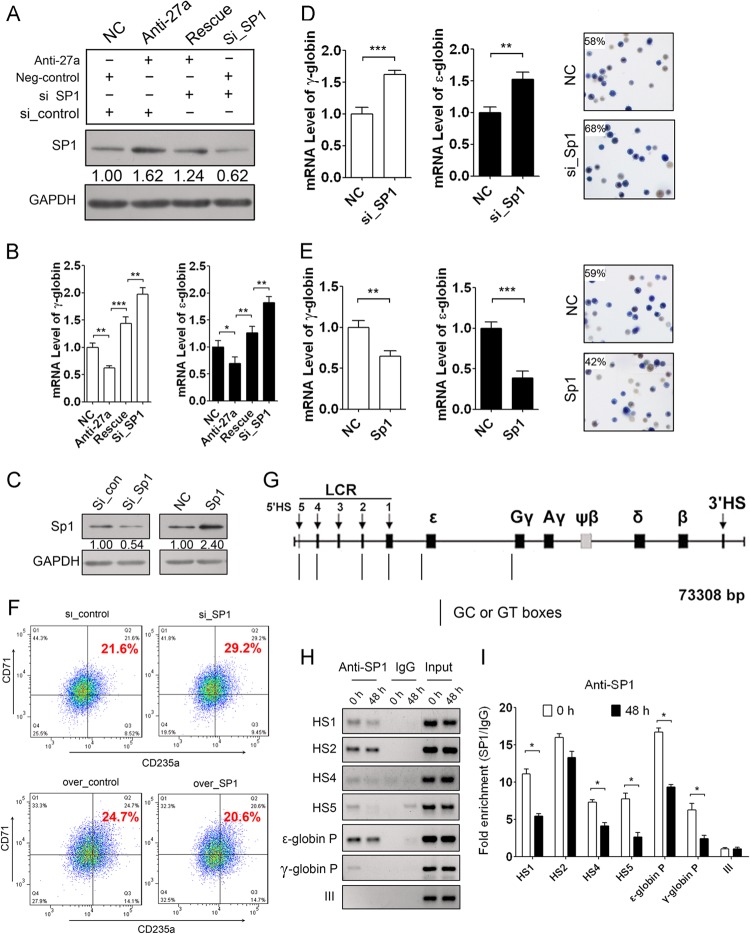
Fig 5.
Repressed SP1 is required for miR-27a-enhanced globin gene expression. (A) Western blot analysis of SP1 in K562 cells cotransfected with miR-27a inhibitor and SP1 siRNA. (B) qPCR analysis of ε- and γ-globin gene expression in the K562 cells shown in panel A. (C) Western blot analysis of SP1 in K562 cells transfected with SP1 siRNA or pCMV-SP1. (D and E) qPCR analysis of ε- and γ-globin gene expression and benzidine staining of hemoglobin-containing cells in K562 cells transfected with SP1 siRNA (D) or pCMV-SP1 (E) at 48 h after hemin induction. (F) FACS analysis of CD235a/CD71 double-positive cells in K562 cells transfected with SP1 siRNA or pCMV-SP1 after 48 h of hemin induction. (G) Schematic of the β-like globin gene locus and SP1 binding sites. (H) ChIP-PCR analysis of the binding of SP1 on the GC or GT boxes in HS1, HS2, HS4, HS5, and the promoter (P) of ε- and γ-globin genes during K562 cell erythroid differentiation. (I) ChIP-qPCR analysis of the binding of SP1 on these sites during K562 cell erythroid differentiation. Student's t test (two-tailed) was performed to analyze the data from the experiments in triplicate. *, P < 0.05; **, P < 0.01; ***, P < 0.001.

