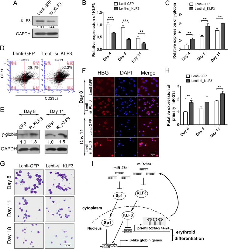
Fig 7.
KLF3 negatively regulates the expression of γ-globin and miR-23a cluster in human primary erythroid cells. (A) Western blot analysis of KLF3 in CD34+ HPCs infected with lentivirus expressing KLF3 shRNA or negative control. (B) qPCR analysis of KLF3 mRNA normalized with GAPDH in CD34+ HPCs infected with lentivirus during Epo-induced erythroid differentiation. (C) qPCR analysis of γ-globin expression normalized with GAPDH in CD34+ HPCs shown in panel B. (D) FACS analysis of CD71/CD235a double-positive cells in CD34+ HPCs infected with lentivirus at 11 days after Epo induction. (E and F) Western blot (E) and immunostaining (F) of γ-globin expression in lentivirus-infected human CD34+ HPCs after 8 and 11 days of Epo induction. (G) May-Grünwald-Giemsa staining of the lentivirus-infected human CD34+ HPCs after 8, 11, and 18 days of Epo induction. (H) qPCR analysis of primary miR-23a cluster in lentivirus-infected human CD34+ HPCs after 4, 8, and 11 days of Epo induction. (I) Schematic of the regulation of miR-23a and miR-27a on β-like globin genes through targeting of KLF3 and SP1. Student's t test (two-tailed) was performed to analyze the data from the experiments in triplicate. **, P < 0.01; ***, P < 0.001.

