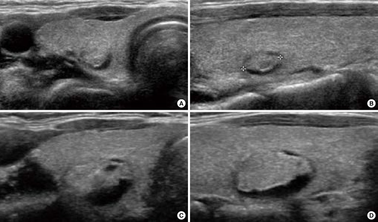
Fig. 1.
Ultrasonography (US) of growing benign mass in a 40-year-old woman who underwent fine needle aspiration twice. (A) Transverse and (B) longitudinal US images demonstrate a 0.7-cm-sized isoechoic nodule with the cystic portion of the posterior portion of the nodule in the right thyroid gland. (C, D) After 4 years, the nodule was enlarged. The cytology was benign, twice.

