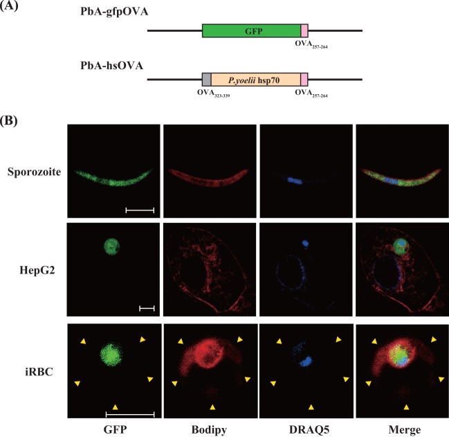
Fig 1.
Expression of a model antigen in the cytoplasm of recombinant P. berghei ANKA. (A) Schematic representation of the transgenic P. berghei ANKA constructs used in this study. (B) PbA-gfpOVA sporozoites, HepG2 cells infected with PbA-gfpOVA sporozoites in vitro, and infected RBCs (iRBC) were stained with BODIPY-TR-C5-ceramide and DRAQ5, which mark membrane structure and nuclei, respectively. Images were obtained using confocal microscopy. Arrowheads, margin of the RBC. Bars, 5 μm.

