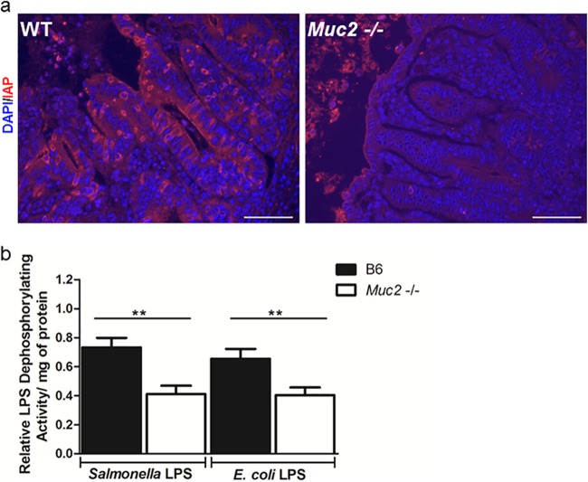
Fig 9.
Muc2−/− mice are impaired in IAP staining and activity. (a) Representative IAP immunostaining (red) in the ceca of WT and Muc2−/− mice at day 7 after infection with ΔaroA S. Typhimurium. The DAPI counterstain is shown in blue. Original magnification, ×400; bars, 20 μm. (b) Analysis of ex vivo LPS dephosphorylation activity in the cecal tissues of WT and Muc2−/− mice infected with ΔaroA S. Typhimurium (assessed at 7 dpi). Homogenized cecal tissues were incubated with Salmonella LPS or Escherichia coli LPS for 2 h, and the malachite green assay was used to measure activity (absorbance at 595 nm). Activity was calculated as relative LPS dephosphorylation activity/mg of protein (normalized to the value for uninfected controls in each group). There were 9 mice per group. Asterisks indicate significant differences (**, P < 0.01) by the Mann-Whitney test.

