
Fig 1.
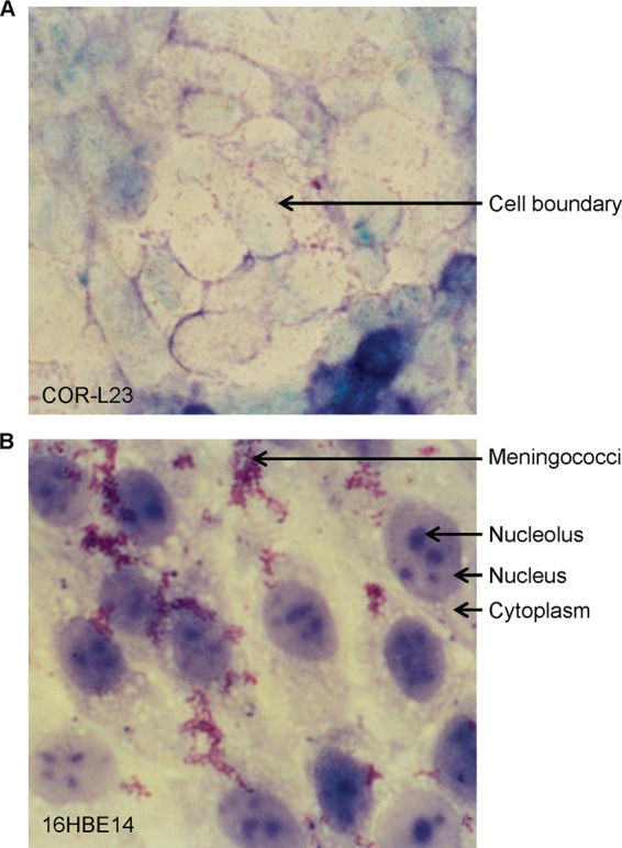
Viability of epithelial cells cocultivated with N. meningitidis MC58. Epithelial cell monolayers cocultivated with bacteria for 48 h were washed and stained with Giemsa. (A) COR-L23 cells at a ×400 magnification. The arrow indicates an example of a cell boundary. (B) 16HBE14 cells at a ×1,000 magnification. Arrows indicate examples of cytoplasm, nucleolus, nucleus, and a group of meningococci.