
Fig 2.
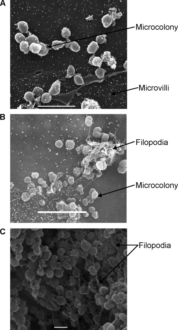
Scanning electron microscopy images of 16HBE14 cells cocultivated with N. meningitidis MC58. Images were taken at 10 h (A), 48 h (B), and 21 days (C) of cocultivation, with white bars indicating 5, 5, and 1 μm, respectively.
Official websites use .gov
A
.gov website belongs to an official
government organization in the United States.
Secure .gov websites use HTTPS
A lock (
) or https:// means you've safely
connected to the .gov website. Share sensitive
information only on official, secure websites.
Scanning electron microscopy images of 16HBE14 cells cocultivated with N. meningitidis MC58. Images were taken at 10 h (A), 48 h (B), and 21 days (C) of cocultivation, with white bars indicating 5, 5, and 1 μm, respectively.