Fig 1.

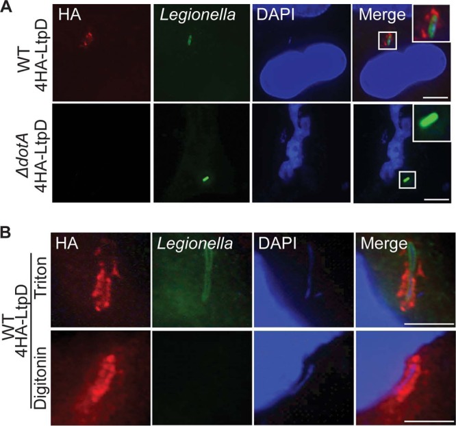
4HA-LtpD localizes to the cytoplasmic surfaces of the LCVs. A549 cells were infected for 5 h with the L. pneumophila 130b WT or ΔdotA mutant expressing 4HA-LtpD, fixed, permeabilized, and immunostained with anti-L. pneumophila (green) and anti-HA (red) antibodies. DNA was visualized with DAPI (blue). (A) After permeabilization with Triton X-100, 4HA-LtpD staining was visible around the WT but not around the ΔdotA bacteria in a manner that suggests the association of 4HA-LtpD with the LCV. (B) After selective permeabilization of membranes with digitonin, 4HA-LtpD staining was detected surrounding the intracellular bacteria, whereas these bacteria were not accessible for anti-L. pneumophila immunostaining. This indicates that 4HA-LtpD is localized on the cytoplasmic surfaces of the LCVs. Images are representative of at least three independent experiments. Scale bars, 10 μm.
