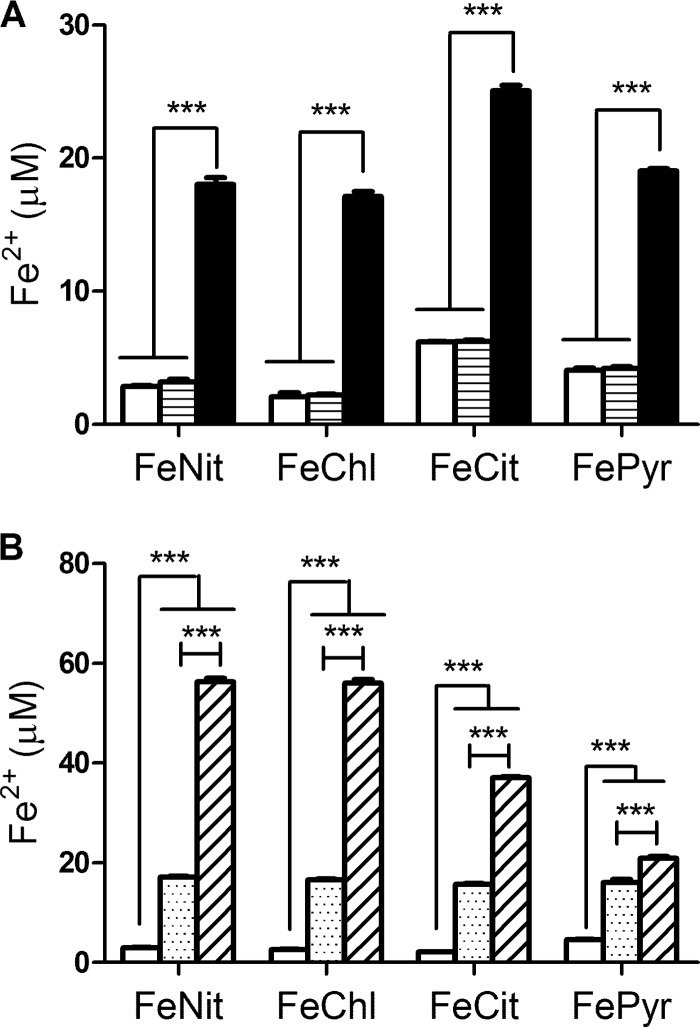
Fig 1.
Reduction of ferric iron by HGA and HGA-melanin. Acid-precipitated HGA-melanin obtained from wild-type strain 130b culture supernatants (black bars) or acid-precipitated material obtained from lly mutant NU408 culture supernatants (bars with horizontal lines) (A), synthetic HGA-melanin (stippled bars) or unpolymerized HGA (bars with diagonal lines) (B), or PBS (white bars) (A, B) was mixed with ferric nitrate (FeNit), ferric chloride (FeChl), ferric ammonium citrate (FeCit), and ferric pyrophosphate (FePyr). After 1 h of incubation for HGA and synthetic HGA-melanin samples or overnight incubation for supernatant samples, the amount of ferrous iron generated was determined. Data are the means and standard deviations obtained from triplicate samples. In panel A, the levels of Fe2+ generated by the wild-type sample were significantly greater than those produced by the mutant sample and the PBS control (***, P < 0.001). In panel B, the levels of Fe2+ generated by HGA were significantly greater than those produced by synthetic melanin (***, P < 0.001), with the results for both HGA and HGA-melanin being greater than those for the PBS control (***, P < 0.001).

