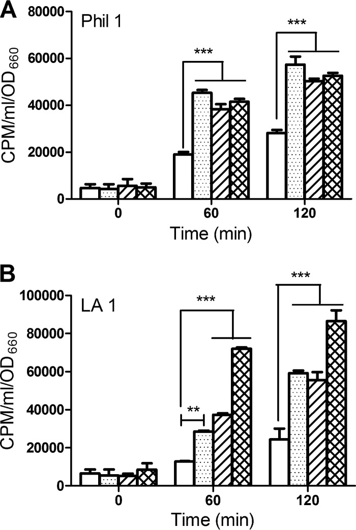
Fig 4.
Iron uptake by other L. pneumophila strains upon exposure to HGA and HGA-melanin. Strains Philadelphia 1 (Phil 1) (A) and Los Angeles 1 (LA 1) (B) that had grown in deferrated CDM were mixed with 55FeCl3 in the presence of either PBS (white bars), synthetic HGA-melanin (stippled bars), unpolymerized HGA (bars with diagonal lines), or vitamin C (hatched bars), and after 0, 60, and 120 min of incubation, the levels of intracellular radiolabeled iron were determined. The data are the means and standard deviations from triplicate samples. In panels A and B, HGA, synthetic HGA, and vitamin C resulted in levels of iron uptake greater than the level for the PBS control (**, P < 0.01; ***, P < 0.001).

