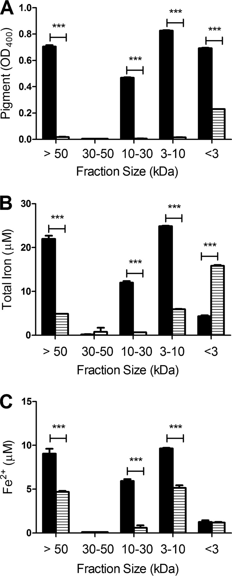
Fig 5.

Association of iron with bacterial HGA-melanin. Wild-type strain 130b (black bars) and lly mutant strain NU408 (bars with horizontal lines) were grown in CDMP supplemented with FeCl3 for 3 days at 37°C, and then cell-free supernatants were collected and consecutively filtered through 50-, 30-, 10-, and 3-kDa cellulose filters. For each fraction, the amount of HGA-melanin was determined by measuring the OD400 of the sample (A), the level of total associated iron was determined by the ferrozine assay in the presence of the reducing agent vitamin C (B), and the amount of associated ferrous iron was ascertained by the ferrozine assay in the absence of vitamin C (C). The data are the means and standard deviations obtained from triplicate samples. In panel A, all wild-type fractions contained more pigment than did the corresponding mutant samples (***, P < 0.001), with the exception of the 30- to 50-kDa fraction, which was devoid of melanin in both cases. In panels B and C, the >50-kDa, 10- to 30-kDa, and 3- to 10-kDa fractions of the wild type contained more iron than did the corresponding fractions of the mutant (***, P < 0.001), whereas the <3-kDa fraction of the mutant had more total iron than that of the wild type (***, P < 0.001).
