Fig 2.
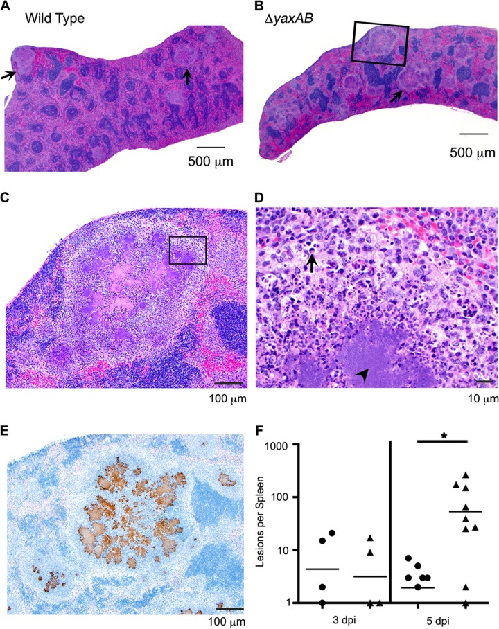
Infection with the ΔyaxAB mutant results in more splenic lesions than wild-type infection. Shown are H&E-stained splenic sections from a mouse infected with either the wild-type (A) or ΔyaxAB (B to D) strain. (A and B) The image at ×2 magnification. Lesions with similar appearance and cellular makeup are present throughout both spleens (examples are highlighted by arrows); however, more lesions are apparent in the ΔyaxAB mutant-infected spleen (B). The region shown in the black box of panel B is enlarged in panel C. (C) The image at ×10 magnification. The region shown in the box is enlarged in panel D. (D) The image at ×60 magnification. The large colonies of bacteria are identified with an arrowhead and are surrounded by numerous neutrophils (identified by segmented or ring-shaped nuclei) and fewer macrophages (identified by round or oval nuclei and abundant foamy cytoplasm). Apoptotic bodies were occasionally observed (arrow). (E) Splenic lesion boxed in panel B stained with anti-Yersinia antibody (brown) showing bacteria localized within the splenic lesions of ΔyaxAB mutant-infected mice. Magnification, ×10. (F) Splenic lesions were counted per spleen in wild-type-infected (●), ΔyaxAB mutant-infected (▲), or mock-infected (not shown) mice. The number of lesions per spleen is shown. Each symbol represents one mouse, and black bars represent the means. At 5 dpi, there are significantly more lesions per spleen in ΔyaxAB mutant-infected mice than wild-type-infected mice, as determined by one-sample t test.

