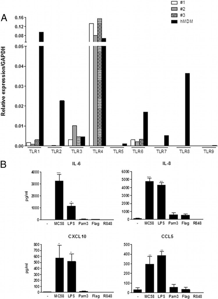
Fig 4.
Meningothelial cell response to TLR stimulation. (A) Expression of TLR by meningothelial cells. Three meningothelial cell lines were analyzed and compared with hMDM for TLR expression determinations. Expression of TLR1, -2, -3, -4, -5, -6, -7, -8, and -9 was determined by QPCR. Data were standardized with reference to GAPDH expression. (B) Secretion of IL-6, IL-8, CXCL10, and CCL5 by meningothelial cells in response to contact with meningococci (strain MC58) or various TLR agonists (n ≥ 4). ANOVA was followed by Dunnett's multiple-comparison test.

