Fig 5.
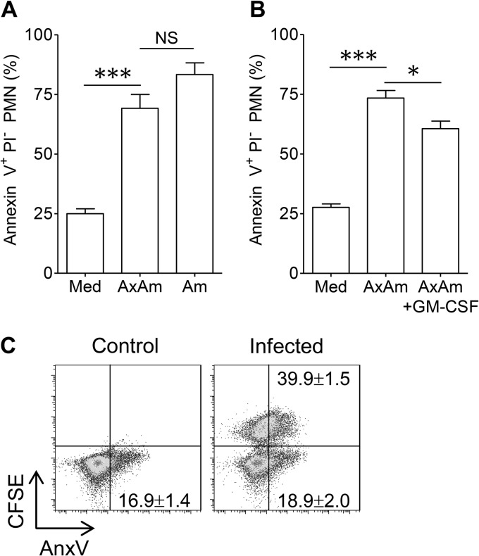
Accelerated neutrophil apoptosis after L. amazonensis amastigote uptake. (A) Percentages of PS+ PI− neutrophils after 18 h of culture in medium (Med) or after coculture with axenic amastigotes (AxAm) or lesion-derived amastigotes (Am). (B) Neutrophil apoptosis in medium alone or in response to axenic amastigotes in the presence or absence of GM-CSF (20 ng/ml). All data in panels A and B are pooled from at least 2 independent repeats and shown as means ± standard errors. NS, not significant. * (P < 0.05) and *** (P < 0.001) indicate statistically significant differences between the groups. (C) Comparison of apoptosis in resting neutrophils, CFSE+ (parasite-carrying) neutrophils, and CFSE− (bystander) neutrophils. PS surface exposure in neutrophils was measured via binding of annexin V (AnxV). The percentages of apoptotic cells were calculated by dividing the number of PS+ cells by the total number of cells for each group. Values are mean percentages of apoptotic cells ± 1 SD. Shown are representative results of one of three independent repeats.

