Fig 1.
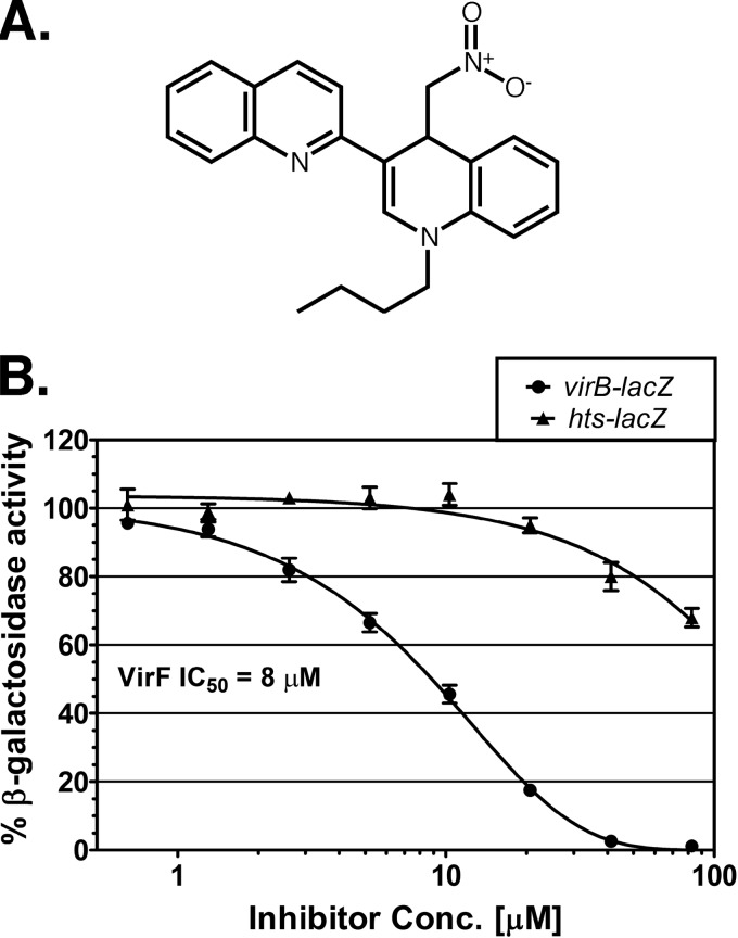
Chemical structure of SE-1 and inhibition of in vivo VirF activation in E. coli. (A) Chemical structure of SE-1 (1-butyl-4-nitromethyl-3-quinolin-2-yl-4H-quinoline). (B) β-Galactosidase (LacZ) activities from two reporter fusions in E. coli, VirF-activated virB-lacZ (SME4382) (circles) and LacI-repressed hts-lacZ (SME3359) (triangles), were assayed at the indicated concentrations of the inhibitor SE-1. VirF and LacI were each expressed from plasmid pHG165. Activity in the absence of SE-1 was set to 100% in each case and corresponded to approximately 950 Miller units for virB-lacZ and 350 Miller units for hts-lacZ. Results are averages for three independent experiments with two replicates each.

