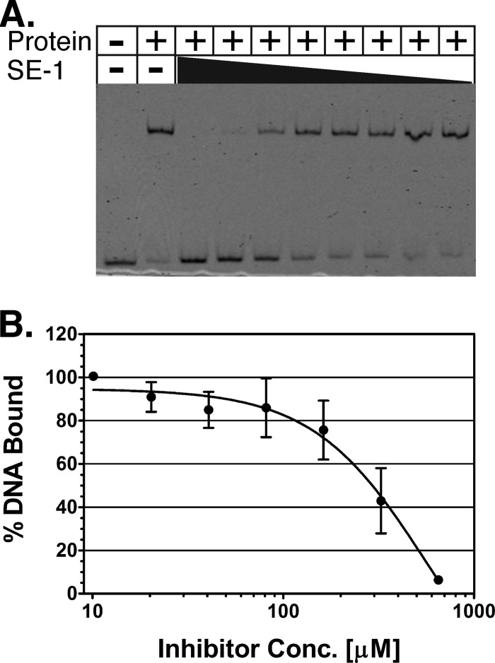
Fig 2.
SE-1 inhibition of in vitro DNA binding by VirF. (A) A representative EMSA gel image. The black triangle represents decreasing concentrations of SE-1, from 1.3 mM to 10 μM, with serial 2-fold dilutions. (B) The binding of purified VirF to a DNA fragment containing the VirF binding site from the virB promoter was assayed using EMSAs with the inhibitor SE-1 at concentrations from 10 to 650 μM (serial 2-fold dilutions). DNA and protein were used at final concentrations of 2 and 300 nM, respectively. The DNA shifted (bound by VirF) was quantified, and the value at the lowest concentration of SE-1 was set to 100%. Results are averages for three independent replicates.

