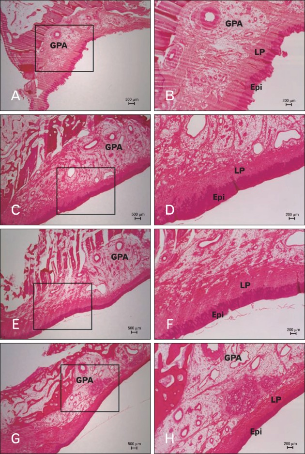
Fig. 3.

Histology sections of the palatal mucosa according to tooth site. (A, B) Canine distal area. (C, D) First premolar distal area. (E, F) Second premolar area. (G, H) First molar distal area. (B), (D), (F), and (H) are boxed areas marked in (A), (C), (E), and (G), respectively. Hematoxylin-eosin stain. Epi, epithelium; GPA, greater palatine artery; LP, lamina propria.
