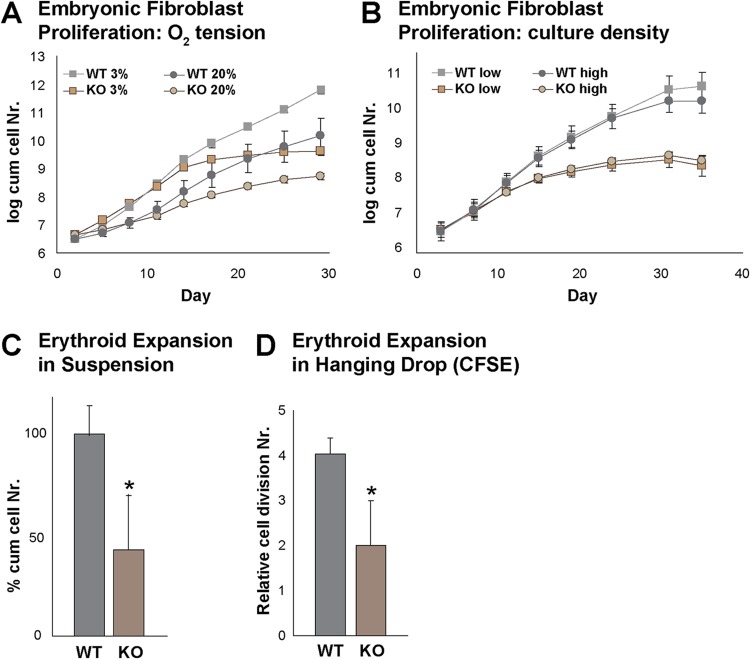
Fig 3.
Proliferation defect in Rad23b-null cells. (A) Proliferation of WT and KO embryonic fibroblasts at 3% and 20% O2 tension. (B) Proliferation of WT and KO embryonic fibroblasts plated at low and high density upon serial passaging at 3% O2 tension. (C) Expansion of WT and KO fetal liver erythroid cells cultured under conditions that favor the proliferation of early erythroid progenitors, preventing their differentiation. (D) Hanging-drop CFSE erythroid differentiation culture showing relative proliferation differences between WT and KO erythroid cells. Asterisks indicate statistical significance.

